Mental Disorders

Clinical Usefulness of IgG Food Allergy Testing

William Shaw Ph.D

Immunoglobulin G (IgG) food allergy testing has made vast advancements since the year 2003 when the American Academy of Allergy, Asthma, and Immunology published a statement that "Measurement of specific IgG antibodies to foods is also unproven as a diagnostic tool"(1) Most of the IgG food allergy throughout the world is done using the same immunochemical technique. First, soluble food proteins in solution are reacted to a solid phase that chemically binds to a variety of proteins. The use of plastic microtiter trays with one to several hundred wells has become the most common material used as the solid phase. Then these trays are washed, dried, and stored for later use. A sample of diluted serum is then added to each of the wells. Antibodies of all types in the diluted serum bind to the specific food molecules that are attached to the plastic wells of the tray. Next, the plates are washed to remove any nonspecific antibodies in the diluted serum. At this time, food antibodies from all of the five major immunoglobulin classes called G, A, M, E, and D may be attached to the food antigens on the plate. The next step confers specificity on the assay. Antisera from sheep, goats, rabbits, or other animals that specifically binds to IgG is added to microtiter wells and only binds to IgG, not to IgA, IgM, IgE, or IgD. This antibody to IgG has previously been modified by the attachment of an enzyme that can be measured conveniently. The amount of enzyme bound to food antigen-IgG complexes on the plate is directly related to how much IgG antibody is attached to a given food. The overall technique is termed Enzyme Linked Immuno Assay or ELISA. If IgG4 is measured, an antiserum specific for IgG4 only must be used for the final step.

The clinical usefulness of IgG testing in an array of illnesses is illustrated in an early article published by an otolaryngologist who reported that the majority of his patients had substantial health improvements after an elimination of foods positive by IgG food allergy tests (2). The overall results demonstrated a 71% success rate for all symptoms achieving at least a 75% improvement level. Of particular interest was the group of patients with chronic, disabling symptoms, unresponsive to other intensive treatments. Whereas 70% obtained 75% or more improvement, 20% of these patients obtained 100% relief. Symptoms most commonly improved 75%-100% on the elimination diets included asthma, coughing, ringing in the ears, chronic fatigue, all types of headaches, gas, bloating, diarrhea, skin rash and itching, and nasal congestion. The most common IgG food allergies were cow's milk, garlic, mustard, egg yolk, tea, and chocolate.

The usefulness of IgG food allergy to design customized elimination diets has now been documented in scientific studies. Irritable bowel syndrome (IBS) is a common, costly, and potentially disabling gastrointestinal (GI) disorder characterized by abdominal pain/discomfort with altered bowel habits (e.g., diarrhea, constipation). The major symptoms of IBS are (1) abnormality of bowel movement, (2) reduction in bowel sensitivity thresholds, and (3) psychological abnormality. Many IBS patients have psychological symptoms including depression, anxiety, tension, insomnia, frustration, hypochondria. psychosocial factors (3). Atkinson et al (4)evaluated a total of 150 outpatients with irritable bowel syndrome (IBS) who were randomized to receive, for three months, either a diet excluding all foods to which they had raised IgG antibodies (ELISA test) or a sham diet excluding the same number of foods but not those to which they had antibodies . Patients on the diet dictated by IgG testing had significantly less symptoms than those on the sham diet after 120 days on the diets. Patients who adhered closely to the diet had a marked improvement in symptoms while those with moderate or low adherence to the IgG test dictated diets had poorer response. Similar results were also obtained by Drisko et al (5). They used both elimination diet and probiotic treatment in an open label study of 20 patients with irritable bowel syndrome diagnosed at a medical school gastroenterology department. The most frequent positive serologic IgG antigen-antibody complexes found on the food IgG tests were: baker's yeast, 17 out of 20 (85%); onion mix, 13 out of 20 (65%); pork, 12 out of 20 (60%); peanut 12 out of 20 (60%); corn, 11 out of 20 (55%);wheat, 10 out of 20 (50%); soybean, 10 (50%); carrot, 9 out of 20 (45%); cheddar cheese, 8 out of 20 (40%); egg white, 8 out of 20 (40%). Only 5 out of 20 reacted by IgG antibody production to dairy; however the majority of patients reported eliminating dairy prior to trial enrollment presumably clearing antigen-antibody complexes prior to testing. Significant improvements were seen in stool frequency, pain, and IBS quality of life scores. Imbalances of beneficial flora and dysbiotic flora were identified in 100% of subjects by comprehensive stool analysis. There was a trend to improvement of beneficial flora after treatment but no change in dysbiotic flora. The one-year follow up demonstrated significant continued adherence to the food rotation diet, minimal symptomatic problems with IBS, and perception of control over IBS. The continued use of probiotics was considered less helpful.

IgG food allergy testing was also proved effective in the gastrointestinal disorder Crohn's disease. Bentz et al (6) found that an elimination diet dictated by IgG food allergy testing resulted in a marked reduction of stool frequency in a double blind cross-over study in which the IgG-dictated diet was compared to a sham diet in 40 patients with Crohn's disease. IgG food allergies were significantly elevated compared to normal controls. Cheese and baker's yeast (Saccharomyces cerevisiae) allergies were extremely common with rates of 83% and 84% respectively. Main et al (7), focusing on the baker's yeast allergy, also found extremely high prevalence of IgG allergy in patients with Crohn's disease. Titers of both IgG and IgA to S. cerevisiae in the patients with Crohn's disease were significantly higher than those in the controls. In contrast, antibody titers in the patients with ulcerative colitis were not significantly different from those in the controls. Among the patients with Crohn's disease there was no significant difference in antibody titers between patients with disease of the small or large bowel. Since IgG antibodies to S. cerevisiae cross react with Candida albicans (8), Candida species colonization might be a trigger for the development of Crohn's disease.

IgG food allergy to wheat, gluten, gliadin, rye, and barley are prevalent in the gastrointestinal disorder celiac disease. Virtually all patients with celiac disease have elevated IgG antibodies to gliadin if they currently have wheat or related grains in their diet. The confirmation of celiac disease is confirmed by the presence of flattened mucosa with a lack of villi when a biopsy sample of the small intestine is examined microscopically. Another confirmation test with equal sensitivity is a blood test for IgA transglutaminase antibodies. The antibody confirmation test is equal in accuracy to the biopsy test with the exception that individuals with IgA deficiency may have false negative results. However, I would estimate that only 1% of people with elevated IgG antibodies to gliadin and other grains related to wheat have celiac disease. If the individual is negative for the confirmation tests for celiac disease, many patients are frequently erroneously advised that that have no problem with wheat. Hadjivassiliou et al argued that it is a significant clinical error to classify wheat allergy through the filter of celiac disease (9) and argue that celiac disease is a subtype of wheat sensitivity. Many of their patients with wheat allergy but celiac disease negative had remission of severe neurological illnesses when they adopted a gluten free diet and expressed that in these patients the gluten molecule causes an autoimmune reaction in the brain rather than in the intestinal tract, likely against the Purkinje cells that are predominant in the cerebellum.

A wide range of additional studies has proven the clinical value of IgG antibodies in autism (10), bipolar depression (11), schizophrenia (12), migraine headaches (13), asthma (14), and obesity (15).

Total IgG Versus IgG4 Food Allergy

Immunoglobulin G (IgG) is classified into several subclasses termed 1, 2, 3, and 4. IgGs are composed of two heavy chain–light chain pairs (half-molecules), which are connected via inter–heavy chain disulfide bonds situated in the hinge region (Figure 1). IgG4 antibodies usually represent less than 6% of the total IgG antibodies. IgG4 antibodies differ functionally from other IgG subclasses in their lack of inflammatory activity, which includes a poor ability to induce complement and immune cell activation because of low affinity for C1q (the q fragment of the first component of complement). Consequently, IgG4 has become the preferred subclass for immunotherapy, in which IgG4 antibodies to antigens are increased to reduce severe antigen reactions mediated by IgE. If antigens preferentially react with IgG4 antibodies, the antigens cannot react with IgE antibodies that might cause anaphylaxis or other severe reactions. Thus, IgG4 antibodies are often termed blocking antibodies. Another property of blood-derived IgG4 is its inability to cross-link identical antigens, which is referred to as "functional monovalency". IgG4 antibodies are dynamic molecules that exchange half of the antibody molecule specific for one antigen with a heavy-light chain pair from another molecule specific for a different antigen, resulting in bi-specific antibodies that are unable to form large cross-linked antibodies that bind complement and thus cause subsequent inflammation(16). In specific immunotherapy with allergen in allergic rhinitis, for example, increases in allergen-specific IgG4 levels indeed correlate with improved clinical responses. IgG4 antibodies not only block IgE mediated food allergies but also block the reactions of food antigens with other IgG subclasses, reducing inflammatory reactions caused by the other IgG subclasses of antibodies to food antigens.

In IgG mediated food allergy testing, the goal is to identify foods that are capable of causing inflammation that can trigger a large number of adverse reactions. IgG1, IgG2, and IgG3 all are capable of causing inflammation because these antibodies do not exchange heavy and light chains with other antibodies to form bispecific antibodies. Thus, IgG1, IgG2, and IgG3 antibodies to food antigens can and do form large immune complexes or lattices that fix complement and increase inflammation. The presence of IgG4 antibodies to food antigens indicates the presence of antibodies to foods that will not usually cause inflammation even though high amounts of these antibodies do indicate the presence of immune reactions against food antigens. Testing only for IgG4 antibodies in foods limits the ability of the clinician to determine those foods that are causing significant clinical reactions that are affecting their patients. The importance of measuring other subtypes of IgG antibodies is highlighted in an article by Kemeny et al. (17). They found that IgG1 antibodies to gluten were elevated in all 20 patients with celiac disease but none of the patients had elevated IgG4 antibodies to gluten.

Clinical References

  • 1. Statement of the AAAAI Work Group Report: Current Approach to the Diagnosis and Management of Adverse Reactions to Foods, October 2003. http://www.aaaai.org/ask-the-expert/usefulness-of-measurements-of-IgG-antibody.aspx (Accessed October 27,2013).

  • 2. Dixon H, Treatment of delayed food allergy based on specific immunoglobulin G RAST testing relief. Otoloryngol Head Neck Surg 2000;123:48-54.

  • 3. Nagisa Sugaya N and Nomura S, Relationship between cognitive appraisals of symptoms and negative mood for subtypes of irritable bowel syndrome. BioPsychoSocial Medicine 2008;2:9-14

  • 4.Atkinson, W et al. Food elimination based on IgG antibodies in irritable bowel syndrome: a randomised controlled trial Gut 2004;53:1459-1464

  • 5. Drisko J, Bischoff B, Hall M, McCallum R, Treating Irritable Bowel Syndrome with a Food Elimination Diet Followed by Food Challenge and Probiotics. Journal of the American College of Nutrition 2006; 25: 514–522

  • 6. Bentz S, et al. Clinical relevance of IgG antibodies against food antigens in Crohn's disease: a double-blind cross-over diet intervention study. Digestion. 2010;81:252-64.

  • 7.Janice Main, Hamish McKenzie, Grant R Yeaman, Michael A Kerr, Deborah Robson, Christopher R Pennington, David Parratt Antibody to Saccharomyces cerevisiae (bakers' yeast) in Crohn's disease BMJ 1988;297:1105-1106

  • 8. Thomas Schaffer, Stefan Mueller, , Beatrice Flogerzi, , Beatrice Seibold-Schmid,Alain M. Schoepfer, and Frank Seibold Anti-Saccharomyces cerevisiae Mannan Antibodies (ASCA) of Crohn's Patients Crossreact with Mannan from Other Yeast Strains, and Murine ASCA IgM Can Be Experimentally Induced with Candida albicans Inflamm Bowel Dis 2007;13:1339 –1346

  • 9. M Hadjivassiliou, R A Grünewald, G A B Davies-Jones Gluten sensitivity as a neurological illness. Neurol Neurosurg Psychiatry 2002;72:560–563

  • 10. Vladimir T et al Higher Plasma Concentration of Food-Specific Antibodies in Persons With Autistic Disorder in Comparison to Their Siblings. Focus Autism Other Dev Disabl 2008; 23: 176-185

  • 11. Severance EG et al Immune activation by casein dietary antigens in bipolar disorder. Bipolar Disord 2010;12: 834–842

  • 12. Severance EG, et al Subunit and whole molecule specificity of the anti-bovine casein immune response in recent onset psychosis and schizophrenia. Schizophr Res. 2010;118:240-7.

  • 13.Huber A, et al Diet restriction in migraine, based on IgG against foods: a clinical double-blind, randomised, cross-over trial. Int Arch Allergy Immunol. 1998; 115:67-72.

  • 14.Vance G. et al. Ovalbumin specific immunoglobulin G and subclass responses through the first five years of life in relation to duration of sensitization and the development of asthma. Clia Exp Allergy 2004;34:1452-1459

  • 15.Wilders-Truschnig M et al. IgG Antibodies Against Food Antigens are Correlated with Inflammation and Intima Media Thickness in Obese Juveniles. Exp Clin Endocrinol Diabetes 2008;116:241-5.

  • 16. Marijn van der Neut Kolfschoten, et al Anti-Inflammatory Activity of Human IgG4 Antibodies by Dynamic Fab Arm Exchange. Science 2007;317:1554-1555

  • 17. Kemeny DM, et al Sub-class of IgG in allergic disease. I. IgG sub-class antibodies in immediate and non-immediate food allergy. Clin Allergy. 1986; 16:571-81.


Vitamin D and Depression

James Greenblatt, M.D.

Vitamin D was thought to be of use only in preventing rickets and osteomalacia. Accumulating evidence, however, has demonstrated that vitamin D does much more, influencing the health and function of tissues and organs throughout the body. Vitamin D is an important nutrient for our physical health, but many people are unaware of how critical this vitamin is for maintaining our mental health. In this article, we will explore the evidence-based research on vitamin D and depression. The next article will explore the effects of vitamin D on psychosis and schizophrenia.

Vitamin D is categorized as a hormone because of its paracrine, autocrine, and endocrine functions, and it can be acquired through food or exposure to the sun. Vitamin D can be found in high amounts in fatty fish and also in milk, yogurt, orange juice and cereals, and dietary supplements. A sufficient amount of vitamin D can also be produced from 5-15 minutes of daily exposure to sunlight.

Vitamin D2, or ergocalciferol, is only made by plants and vitamin D3, or cholecalciferol, is created when ultraviolet light hitting the skin photochemically converts cholesterol to vitamin D. Serum 25-hydroxyvitamin D [25(OH)D] is a biological marker used to reliably measure the levels of both forms of vitamin D. Among its many functions, vitamin D is important for absorbing calcium, maintaining calcium homeostasis in tissues, growth of bones and teeth, properly functioning neurons and glial cells, preventing rickets and osteomalacia, influencing tissues and organs, preventing psoriasis, muscle pain, weakness, elevated blood pressure, some forms of cancer, and autoimmune disease, and preserving mental health.

Recent studies have advanced our understanding of vitamin D and its effect on the brain. There are vitamin D receptors in neurons and glial cells in the brain. Specifically, research suggests vitamin D may act on particular regions of the brain important in the development of depression, including the prefrontal cortex, hippocampus, cingulate gyrus, thalamus, hypothalamus, and substantia nigra. Moreover, it has also been discovered that genetic variations of vitamin D receptors are associated with depression. Recent research shows vitamin D controls the transcription of over one thousand genes involved in neurotrophic and neuroprotective effects, including the maintenance and development of neurons. In addition, vitamin D may also stimulate the release of neurotrophins, a family of proteins that function to protect and stimulate the growth of neurons.

In a recent study, Polak et al. investigated the association between vitamin D levels and depressive symptoms in 615 young adults. Subjects in the lowest quartile of vitamin D levels were more likely to report having symptoms of depression than those in the highest quartile, suggesting that vitamin D deficiency is a potential predictor of depression. Similarly, in a study on previously deployed military personnel who committed suicide, Umhau et al. found that subjects in the lowest octile of vitamin D levels had the highest risk of suicide. Milaneschi et al. found a comparable effect in the elderly population, with low levels of vitamin D correlating with a significantly higher risk of developing depression. In another study with adolescent participants, Toppanen et al. measured vitamin D levels and depressive symptoms in the same group of children at 9.8, 10.6, and 13.8 years old. Interestingly, higher levels of vitamin D at age 9.8 predicted lower levels of depression at age 13.8, suggesting an association between low levels of vitamin D and early onset depression.

Bertone-Johnson et al., performed a cross-sectional study on 81,189 older women and found an inverse association between vitamin D levels and depressive symptoms in the postmenopausal women. In another study, Lee et al. found that lower vitamin D levels were associated with depression in a population of 3,369 European men. A study by Black et al. came to the same results in a population of young adult males.

Vitamin D supplements have also been found to enhance positive moods. In a study by Allen et al., healthy subjects were given 800 IU, 400 IU, or no vitamin D during five days of winter. The results of their study showed that vitamin D was able to significantly enhance positive affect and also reduce negative affect. Taken together, these diverse studies suggest an indisputable connection between vitamin D deficiency and depression across all age groups and genders.

Biochemical individuality plays a substantial role in vitamin D status. Although environmental factors, such as nutrition and sun exposure, are considered the major determinants of vitamin D status, genetics are responsible for a large portion of the variation seen in serum 25-hydroxyvitamin D. A Swedish study involving 204 same-sex twins between the ages of thirty-nine and eighty-five years living at northern latitude 60 degrees found that genetic factors were responsible for one-fourth of the variation in serum 25-hydroxyvitamin D, independent of season. During the summer season alone, genetics was responsible for half of the variability in 25-hydroxyvitamin D.

Vitamin D levels between 20 and 30 ng/mL have been traditionally accepted as normal and healthy. We now know that this range is too low, and even people who were thought to be safely in the middle of range may need vitamin D supplementation. Given the quirks of biochemical individuality, some people in the upper reaches may need even more. I prefer to see a 25-hydroxyvitamin D between 40 and 60 ng/mL in my patients. The best way to determine vitamin D deficiency is through serum blood testing which should also be done twice a year.

Research literature supports a link between vitamin D and depression; however, the exact mechanisms are unclear. The research has not yet established whether low levels of vitamin D cause depression, or whether depression causes low levels of vitamin D. New research is continually emerging on the importance of vitamin D in sustaining mental health. In one 2012 study, adolescents in a mental health facility who were vitamin D deficient were 3½ times more likely to have psychotic features when compared to those with sufficient vitamin D levels. We'll explore the exciting research implicating vitamin D's role in other mental illnesses such as psychosis and schizophrenia in the next newsletter.

Clinical References


  • Anglin R, Samaan Z, Walter S et al. Vitamin D deficiency and depression in adults: systematic review and meta analysis. British Journal of Psychiatry, 2013.

  • Bertone-Johnson ER, Powers SI, Spangler L et al. Vitamin D Supplementation and Depression in the Women's Health Initiative Calcium and Vitamin D Trial. Am J Epidemiol 2012; 176(1):1-13.

  • Bertone-Johnson ER, Powers SI, Spangler L, et al. Vitamin D intake from foods and supplements and depressive symptoms in a diverse population of older women. Am J Clin Nutr. 2011 Oct;94(4):1104-12.

  • Bertone-Johnson ER. Vitamin D and the occurrence of depression: causal association or circumstantial evidence? Nutr Rev. 2009 Aug;67(8):481-92.

  • Black LJ, Jacoby P, Allen KL, et al. Low vitamin D levels are associated with symptoms of depression in young adult males. Aust N Z J Psychiatry 2014 May;48(5):464-71.

  • Dean A J, Bellgrove M A, Hall T et al. Effects of vitamin D supplementation on cognitive and emotional functioning in young adults–a randomised controlled trial. PLoS One. 2011;6(11):e25966

  • Gracious, BL, Finucane, TL, Friedman-Campbell, M. et al. Vitamin D deficiency and psychotic features in mentally ill adolescents: A cross-sectional study. BMC Psychiatr. 2012; 12: 38.

  • Han B, Lyu Y, Sun Y et al. Low serum levels of vitamin D are associated with post-stroke depression. European Journal of Neurology Dec 2014. [E-pub ahead of print].

  • Jorde, M. Sneve, Y. Figenschau, J et al. Effects of vitamin D supplementation on symptoms of depression in overweight and obese subjects: randomized double blind trial. J Intern Med. 2008;264(6):599-609.

  • Lansdowne AT & Provost SC. Vitamin D3 enhances mood in healthy subjects during winter. Psychopharmacology (Berl) 1998 Feb;135(4):319-23

  • Lee DM, Tajar A, O'Neill TW, et al. Lower vitamin D levels are associated with depression among community-dwelling European men. J Psychopharmacol 2011 Oct;25(10):1320-8.

  • Polak MA, Houghton LA, Reeder AI, et al. Serum 25-hydroxyvitamin D concentrations and depressive symptoms among young adult men and women. Nutrients 2014 Oct 28;6(11):4720-30.

  • Toffanello ED, Sergi G, Veronese N, et al. Serum 25-hydroxyvitamin d and the onset of late-life depressive mood in older men and women: the Pro.V.A. study. J Gerontol A Biol Sci Med Sci 2014 Dec;69(12):1554-61.

  • Tolppanen AM, Sayers A, Fraser WD, et al. The association of serum 25-hydroxyvitamin D3 and D2 with depressive symptoms in childhood--a prospective cohort study. J Child Psychol Psychiatry 2012 Jul;53(7):757-66.

  • Umhau JC, George DT, Heaney RP, et al. Low Vitamin D Status and Suicide: A Case-Control Study of Active Duty Military Service Members. PLoS One 2013;8(1):e51543.

  • Yue W, Xiang L, Zhang YJ, et al. Association of serum 25-hydroxyvitamin D with symptoms of depression after 6 months in stroke patients. Neurochem Res. 2014 Nov;39(11):2218-24.

Candida: A Factor in Depression and Mental Health

Jessica Bonovich, R.N.

One of the first psychiatrists to publish findings about the connection between Candida and depression is Dr. Orian Truss. His compelling work has been cited in numerous books and helped countless patients to date. Interestingly, his discovery in 1981 did not generate much interest from the psychiatric community who had just begun to see the effects of MAO inhibitors. While these and other modern antidepressants remain an important tool for treating psychiatric patients, studies have demonstrated that their efficacy rate is only about 20-30 percent (Kroenke, Hansen). Clearly, we do not have sufficient understanding of the complex spectrum of mental disorders that plague millions of individuals. To this end, we should leave no stone unturned. Especially if that stone has already shown to provide helpful information.

Dr. Truss hypothesized that where Candida is merely a nuisance for some, it causes chronic illness (including mental illness) in others. People with weakened immune systems are particularly at risk however they need not have HIV or leukemia to suffer. At the Huxley symposium (1981), he presented 6 case studies he believed to encompass the so-called Candida syndrome that he discovered. All individuals had been exposed to multiple rounds of antibiotics or other immune lowering agents. These individuals were often female. Depression was almost always one of many vague symptoms. Loss of memory, difficulty concentrating, sensitivity to chemicals and other symptoms were also noted. Dr. Truss began to incorporate treatment with antifungal therapy for patients with chronic mental illness. Interestingly, all of the individuals responded to the treatment and their symptoms of depression lifted.

More recently, a double blind placebo controlled study in 2001, under the direction of Heiko Santelmann found the antifungal drug nystatin to be significantly more effective at reducing symptoms of depression in polysymptomatic patients. In fact, the authors of the study noted that some of the most dramatic improvements reported were from individuals who had mental complaints. While nystatin is not specific to Candida, it is a compelling study that demonstrates yeast can affect mental health.

Dr. Truss noted the difficultly of testing for Candida since nearly everyone has had exposure to the organism. Determining the degree to which each individual suffers is based on a set of vague symptoms that often ends with the patient being labeled as psychosomatic or psychiatric. Fortunately, today we have more robust methods of detecting Candida and a better understanding of the mechanism which may be causing symptoms.

Genetic coding has helped determine the mechanisms that pathogens employ to help increase virulence. In the case of Candida albicans, a specific gene that codes for Immunogenic Alcohol Dehydrogenase was detailed in 1994 (Bertryam). This enzyme produces acetylaldehyde from glucose or ethanol (Gainza-Cirauqui). This acetylaldehyde creates an environment that is not conducive to most microbes which effectively decreases the competition. In humans, acetylaldehyde is a carcinogenic compound that easily passes the blood brain barrier where it interferes with neurotransmission (Correa). Depression and acetylaldehyde both cause a reduction in natural killer cell cytotoxicity (Irwin). Since nearly everyone has Candida in the body, it is plausible that a brief episode of depression may increase the possibility of developing a chronic condition.

For years, a yeast culture with sensitivity has been the mainstay of Candida testing. The benefit to this test is that it can determine the exact species of yeast so that appropriate treatment can be instituted. The problem with this method is that it is notorious for producing false negatives (Maaroufi). Metabolites of yeast detected in The Great Plains Laboratory Organic Acids Test are a very reliable method of detecting Candida overgrowth (Shaw). While this test cannot determine the exact organism, this is less important. Most prescriptive antifungal agents are effective at killing Candida albicans, which is the most common yeast species (Shaw).

Armed with correct information, the latest in diagnostic testing, and viable treatment options, psychiatric and primary care physicians can exercise multiple options for patients with symptoms of depression and other mental health disorders. As more and more physicians are looking outside the box to find solutions to psychiatric diseases as complex as the patients themselves, many are turning to comprehensive testing for yeast (and other pathogens) for answers. If a patient presents with a recent onset of chronic depression in the absence of major trauma, it makes sense to ask the question: Could this be related to Candida or another pathogen? Doing so may save your patient's life, or at least the life they once knew!

Clinical References

  • Hansen, R. et al. (2005). Efficacy and Safety of Second-Generation Antidepressants in the Treatment of Major Depressive Disorder. Annals of Internal Medicine, 143(6); 415-426.

  • Kroenke, K., et al. (2001). Similar Effectiveness of Paroxetine, Fluoetine, and Sertaline in Primary Care. JAMA, 286(23); 2947-2955

  • Truss, O. (1981). The Role of Candida in Human Illness. Presented that the Huxley symposium, September, Birmingham, AL.

  • Bertryam, G., et al. (1995). Structure and Regulation of the Candida albicans ADH1 Gene Encoding an Immunogenic Alcohol Dehydrogenase. Yeast, 12:115-127.

  • Santelmann, H et al. (2001). Effectiveness of nystatin in polysymptomatic patients. A randomized, double-blind trial with nystatin versus placebo in general practice. Family Practice, 18; 258-265.

  • Gainza-Cirauqui, ML., et al. (2013). Production of carcinogenic acetaldehyde by Candida albicans from patients with potentially malignant oral mucosal disorders. Journal of Oral Pathology and Medicine, 42(3); 243-9.

  • Correa, M., et al. (2011). Piecing together the puzzle of acetaldehyde as a neuroactive agent. Neuroscience and Biobehavioral Reviews, 36; 404-430.

  • Irwin, M., et al. (1990). Major Depressive Disorder, Alcoholism, and Reduced Natural Killer Cell Cytotoxicity: Role of Severity of Depressive Symptoms and Alcohol Consumption. JAMA Psychiatry, 47(8); 713-719.

  • Maaroufi, Y., Heymans, C., De Rune, J., Duchateau, H. (2003). Rapid Detection of Candida albicans in Clinical Blood Samples by Using a TaqMan-Based PCR Assay. Journal of Clinical Microbiology, 41; 3293-3298.

  • Shaw ,W., (2008) Biological Treatments for Autism and PDD. Publisher: Author.

Rickets and eye-poking in autism associated with calcium deficiency

William Shaw Ph.D

I strongly disagree with the assertion by some individuals in the autism field that calcium is a problem to individuals on the autistic spectrum. Failure to provide adequate calcium is very dangerous and could lead to the loss of the eyes due to severe eye-poking behavior. Calcium deficiency can be a severe problem in normal children on a milk free diet since milk is a significant source of protein, vitamin D, and calcium needed for strong bones and teeth. Some physicians have reported rickets (1), a severe bone deformity, occurs in children with autism on the gluten and casein free diet who did not receive added calcium supplements. Calcium and vitamin D supplementation is essential to children on a casein free diet since most children with autism do not eat substantial amounts of other calcium-rich foods. Use of milk substitutes like B-Unique® provides adequate calcium, protein, and fat comparable to whole milk without the presence of casein and lactose that are problematic in most children with autism.

Children with autism may have an even more severe problem with calcium deficiency. Mary Coleman, M.D. (2) reported that children with autism who are calcium deficient are much more likely to poke out their eyes and a substantial number of children with autism have done so. I have talked to numerous parents of children with autism that began to touch their eyes after starting the casein-free diet. This abnormal behavior is associated with low urine calcium; blood calcium levels were usually normal. Treatment with calcium supplementation prevents this behavior. (I suspect that this behavior is due to increased eye pain due to high substance P or to deposits of oxalate crystals in the eye. Low calcium may act to intensify this pain and poking out the eye relieves the pain.) Dr. Coleman also found that speech developed very quickly after calcium supplementation in a portion of mute children with autism who had low urine calcium. Parathyroid hormone, calcitonin, and vitamin D were all normal in patients with autism but all of them had low urine calcium. In one case, according to a parent who contacted me, her child with autism persisted in poking at the eyes even after one eye had been poked out and surgically replaced. Calcium supplementation stopped this behavior immediately. I am aware of many other children with eye-poking behavior in which calcium supplements stopped this behavior in less than two days. Verbal autistic children say that their eye pain is severe and that calcium supplementation stopped their pain quickly. The urine calcium and magnesium tests will soon be a part of the Organic Acids Test offered by The Great Plains Laboratory, Inc.

It is important that calcium, magnesium, and zinc be in balance for optimal nutrition. Vitamin D supplementation may also be needed when milk is eliminated unless other sources of vitamin D are included in the diet or the child is exposed to adequate sunlight. Children with autism also need additional calcium to prevent oxalate deposition in the tissues. Although sardines and dark leafy greens like spinach, kale, turnips, and collard greens are high in calcium, all of these foods except sardines are high in oxalates. High oxalates can be fatal if formed in the renal tract. Oxalates in the urine are much higher in individuals with autism than in normal children. As a matter of fact, 36% of the children on the autistic spectrum had values higher than 90 mmol/mol creatinine, the value consistent with a diagnosis of genetic hyperoxalurias while none of the normal children had values this high. 84% of the children on the autistic spectrum had oxalate values outside the normal range (mean ± 2 std dev). None of the children on the autistic spectrum had elevations of the other organic acids (glyceric and glycolic acids) associated with genetic diseases of oxalate metabolism, indicating that oxalates are high due to external sources. When calcium is taken with foods that are high in oxalates, oxalic acid in the intestine combines with calcium to form insoluble calcium oxalate crystals that are eliminated in the stool. This form of oxalate cannot be absorbed into the body. When calcium is low in the diet, oxalic acid is soluble in the liquid portion of the contents of the intestine (called chyme) and is readily absorbed from the intestine into the bloodstream. If oxalic acid is very high in the blood being filtered by the kidney, it may combine with calcium to form crystals that may block urine flow and cause severe pain. However, such crystals may also form in the bones, joints, blood vessels, lungs, eyes, skin, heart, thymus, skeletal muscle, joints, fat, teeth, mouth, nerves, and even the brain. In addition, oxalate crystals in the bone may crowd out the bone marrow cells, leading to anemia and immunosuppression. Calcium citrate is the best source of calcium to prevent oxalate absorption because citrate ion blocks the absorption of oxalates in the intestinal tract (3). A combination of calcium citrate and magnesium citrate is the best form of supplement to provide calcium and magnesium needs while preventing excess oxalate buildups in the body.

Clinical References:

  • 1. Hediger ML, England LJ,Molloy CA, Yu KF, Manning-Courtney P, Mills JL. Reduced bone cortical thickness in boys with autism or autism spectrum disorder. J Autism Dev Disord. 2008;38(5):848–856

  • 2. Coleman, M. Clinical presentations of patients with autism and hypocalcinuria. Develop. Brain Dys. 7: 63-70, 1994

  • 3. Caudarella R, Vescini F, Buffa A, Stefoni S. Citrate and mineral metabolism: kidney stones and bone disease. Front Biosci. 2003 Sep 1;8:s1084-106.

The Implications of Low Cholesterol in Depression and Suicide

James M. Greenblatt, M.D.

For the last quarter century, we have been told that cholesterol is dangerous for our health and were advised to avoid it in order to live a healthier life. However, cholesterol is essential in maintaining good mental health. The brain is the most cholesterol-rich organ in the body, and depriving the brain of essential fatty acids and cholesterol can lead to detrimental health problems. Lower levels of cholesterol in the blood are associated with a heightened risk of developing major depressive disorder, as well as an increased risk of death from suicide. A study published in the Journal of Psychiatric Research found that depressed men with low total cholesterol levels (less than 165 milligrams per deciliter [mg/dL]) were seven times more likely to die prematurely from unnatural causes such as suicide and accidents.

Most recently, the continued allegation that cholesterol is dangerous came under scrutiny. A meta-analysis published in the March 2014 issue of Annals of Internal Medicine found that there's not enough evidence supporting the claim that saturated fat increases the risk of heart disease. After reviewing 72 different studies, researchers did not find that people who ate higher levels of saturated fat had more heart disease than those who ate less. Researchers came to the conclusion that instead of avoiding fats, which are essential to maintaining brain health, scientists are identifying the real villains as sugar and highly processed foods.

Low Cholesterol and Depression

Several studies have linked low cholesterol levels to an increased risk of developing depression. Consider the following examples:

  • A 1993 paper published in the Lancet reported, "Among men aged seventy years and older, categorically defined depression was three times more common in the group with low total plasma cholesterol . . . than in those with higher concentrations."

  • A 2000 study published in Psychosomatic Medicine, researchers compared cholesterol levels to depressive symptoms in men ranging in age from forty to seventy. They found that men with long-term, low total cholesterol levels "have a higher prevalence of depressive symptoms" compared to those with higher cholesterol levels.

  • Women with low cholesterol levels are also vulnerable to depression. In 1998, Swedish researchers reported the results of their examination of cholesterol and depressive symptoms among 300 healthy women, ages thirty-one to sixty-five, in and around Stockholm. Women in the lowest cholesterol group (the bottom tenth percentile) suffered from significantly more depressive symptoms than did the others.

  • A 2001 study published in Psychiatry Research looked at primary care patients in Ireland, finding that low levels of cholesterol were linked to higher ratings on depression rating scales.

  • Italian researchers measured the cholesterol levels of 186 patients hospitalized for depression and found an association between low cholesterol and depressive symptoms.

This research is supported by other studies, including a 2008 meta-analysis, which found that higher total cholesterol was associated with lower levels of depression. A 2010 study published in The Journal of Neuropsychiatry & Clinical Neurosciences looked at the levels of HDL in depressed people and found that low levels of HDL were linked to "long-term depressive symptomatology."

Low Cholesterol and Suicide

Suffering through a depressive episode can be very difficult, and one of the great fears is that someone in the throes of depression does not see any point in continuing to live.

Early evidence of a link between low cholesterol and suicide came from the Multiple Risk Factor Intervention Trial study, a large-scale, long- term look at various health factors involving hundreds of thousands of volunteers. Data from the study was analyzed by researchers from the University of Minnesota, who found that people with total cholesterol levels lower than 160 mg/dL were more likely to commit suicide than those with higher cholesterol levels. Other studies are equally alarming:

  • A 2008 study looked at forty men who were hospitalized due to bipolar disorder. Twenty had attempted suicide at some point in the past, and the other twenty had not. Both cholesterol and blood fat levels were lower, on average, among those who had attempted suicide.

  • A paper published in the Journal of Clinical Psychiatry in the same year reported the results of an examination of cholesterol levels in 417 patients who had attempted suicide at some point, 155 hospitalized psychiatric patients who had not, and healthy controls. Results of the study suggest that low cholesterol may be associated with suicide attempts.

  • The suicidal method of choice, self-inflicted fatal gun wound versus pills, for example, may also be related to cholesterol levels. A2008 study published in Psychiatry Research compared nineteen people who had attempted suicide using violent methods to sixteen who had attempted to kill themselves nonviolently, as well as to twenty healthy controls. The researchers found that "violent suicide attempters had significantly lower total cholesterol and leptin levels compared with those with nonviolent suicide attempts."

The connection between low cholesterol and suicide is highlighted in a 2004 study, which concluded that a low total cholesterol level can be used as an indicator of suicide risk. This study, involving suicide attempters with major depressive disorder, nonsuicidal depressed patients, and normal controls, found significant differences in cholesterol levels among the various groups.

The average total serum cholesterol level was 190 mg/dL among the normal controls, 180 mg/dL in nonsuicidal depressed group, and 150 mg/dL among the suicidal depressive patients. This study showed that the total cholesterol level can be used to gauge possible suicide risk (less than 180 mg/dL) and probable risk (150 mg/dL and lower).

Suicide is not the only type of violence associated with lower cholesterol levels. Homicide and other violence committed against others is also associated with low cholesterol. Swedish researchers compared one-time cholesterol measurements on nearly eighty thousand men and women, ranging in age from twenty-four to seventy, to subsequent arrests for violent crime. The researchers reported that "low cholesterol is associated with increased subsequent criminal violence."

What's the Cholesterol-Depression Link?

There is strong scientific evidence indicating that low cholesterol and suicide, particularly violent suicide, are linked. The vast majority of studies linking low cholesterol to depression, suicide, and violence looked at the serum cholesterol level. But what about the amount of cholesterol in the brain?

Canadian researchers were the first to examine this question in their 2007 study published in the International Journal of Neuropsychopharmacology. The researchers measured and compared the cholesterol content in various parts of the brains of forty-one men who had committed suicide and twenty-one men who had died of other, sudden causes that had no direct impact on the brain. The results were intriguing: When the suicides were categorized as violent or nonviolent, those who had committed violent suicide were found to have less cholesterol than the others in the gray matter of their brains. This was seen specifically in the frontal cortex, a part of the brain that handles "executive functions," including processes involved in planning, cognitive flexibility, abstract thinking, initiating appropriate actions and inhibiting inappropriate actions, and selecting relevant sensory information. The frontal cortex essentially controls the ability to make good decisions.

Cholesterol is a critical precursor to many essential physiological molecules in the human body that directly and indirectly affect our moods and optimal brain function. Some researchers theorize that low levels of cholesterol alter brain chemistry, suppressing the production and/or availability of the neurotransmitter serotonin. Cholesterol is essential for the synthesis of all steroid and sex hormones, including DHEA, testosterone, and estrogen. Cholesterol is also needed in the synthesis of vitamin D.

Clinically low cholesterol is a significant variable in the treatment and recovery from mood disorders. A simple blood test looking at total cholesterol can reflect multiple factors influencing treatment. In my clinical practice for the past 20 years, I have found that low cholesterol (<130) has significant implications for what is referred to as "treatment-refractory" depression. This refers to patients who have failed to recover from traditional antidepressant medications. Treatment-refractory patients often struggle with intense suicidal ideation and aggressive behavior. Often, we are able to determine that low cholesterol is genetic, as there are other members in the family who also have low cholesterol levels, despite eating a diet rich in cholesterol and saturated fats. For individuals with low cholesterol, a diet with adequate cholesterol and saturated fats is highly recommended in order to replenish cholesterol levels, although supplemental cholesterol may also be needed for many.

New Beginning's Sonic Cholesterol supplement provides 250 milligrams of cholesterol per capsule. Individuals with low cholesterol levels may take between two to six capsules per day in order to restore adequate cholesterol levels for optimal brain function. Cholesterol repletion is often slow and can take many months. Once cholesterol levels are normalized, we often see an improvement in symptoms and a decreased dependency on medications. It is quite striking to consistently witness the high correlation between cholesterol levels and behavioral and mood symptoms.

For information about cholesterol supplementation, contact New Beginnings Nutritionals.

Conclusion

There is a growing amount of research looking at the use of essential fatty acids, particularly omega-3's in psychiatry, but we often overlook cholesterol. Low levels of cholesterol and essential fatty acids are intimately linked to depression. Understanding the consequences of deficiencies in essential fats and cholesterol is important for the effective treatment of depression. Whether it is drug induced, genetic, or a result of dietary patterns, low cholesterol impairs optimal brain function and often prevents successful recovery from chronic depression.

Clinical References:

  • 1. Hediger ML, England LJ,Molloy CA, Yu KF, Manning-Courtney P, Mills JL. Reduced bone cortical thickness in boys with autism or autism spectrum disorder. J Autism Dev Disord. 2008;38(5):848–856

  • 2. Coleman, M. Clinical presentations of patients with autism and hypocalcinuria. Develop. Brain Dys. 7: 63-70, 1994

  • 3. Caudarella R, Vescini F, Buffa A, Stefoni S. Citrate and mineral metabolism: kidney stones and bone disease. Front Biosci. 2003 Sep 1;8:s1084-106.

Evidence that Increased Acetaminophen use in Genetically Vulnerable Children Appears to be a Major Cause of the Epidemics of Autism, Attention Deficit with Hyperactivity, and Asthma

William Shaw, Ph.D.

Introduction

One of the puzzling aspects of autism is the marked increase in the incidence of autism that began in the United States in the early 1980s and has appeared to increase continuously since then. The highest incidence of autism has been reported to be South Korea, where the incidence is now reported to be one in 38 boys. [1]Increased incidence of autism due to more effective diagnosis was disproved in the study of Irva Hertz-Picciotto who showed that perhaps 12% of the increased autism diagnoses could be attributed to improved diagnosis [2]. A wide range of environmental factors has been associated with increased autism incidence, including pesticides, chemicals, phthalates, polychlorinated biphenyls, solvents, heavy metals or other pollutants. [3]Although toxic chemicals are undoubtedly not beneficial for the health of any person, is there any information that indicates that a toxic avalanche of chemicals inundated the United States in the early 1980s? Indeed a wealth of knowledge about environmental chemicals has led to marked reductions in exposure to chemicals such as lead and dichlorodiphenyltrichloroethane (DDT) in the United States over the past 50 years. For example, acceptable safe limits for levels of lead in the blood have decreased from 60 μg/dL in 1960 to <5 μg/dL in 2010 [4].

Making a connection between disease appearance and causative agent is important. Clinical studies, epidemiological studies and post-market pharmacovigilence are of utmost importance in recognizing signals and drug-induced side effects. One of the most notable cases of serious adverse effects caused by a pharmaceutical agent was the terrible developmental epidemic of the birth of children with seal-like arms and legs (phocomelia) that was linked to the maternal use of the sedative thalidomide 20–35 days after conception [5]. What would have happened if the thalidomide connection had never been made? One of the difficulties with chemical studies of autism associations is that most chemicals are used worldwide, making it difficult to find a "clean" environment where autism might be less prevalent. One of the clues that led to the discovery of thalidomide as the causative agent of deformed limbs was that it was much more commonly used in Europe than in the United States. Countries with the greatest use of thalidomide by pregnant women during pregnancy were those with the highest incidence of deformed babies. If there was a geographic region in the world in which the incidence of autism was much lower than that in the United States, a comparison of medical or dietary differences might provide a significant clue to the major cause of autism.

Such a country is Cuba. The highest estimate of the total incidence of autism in Cuba is 185 cases out of a total population of 11,000,000 (0.00168% of the population) compared with an estimate of as high as 1.5 million in a total United States population of 300 million (0.50%) [6, 7]. The percentage of the population with autism in the United States is thus 298 times higher than in Cuba. Cuba is much more economically challenged than the United States, with the per capita income of Cuba approximately eight times lower than that in the United States. Despite the economic challenges presented to the communist government of Cuba, basic healthcare is readily available and there are a large number of physicians trained in 14 different medical schools. Unlike the United States, where vaccines are optional in many states, vaccines are compulsory in Cuba and Cuba has one of the most highly vaccinated populations in the world against a wide variety of infectious agents.[8] For example, the vaccination rate for measles was reported to be 99.7%. The association of autism with various vaccines has had a very controversial history with inflamed passions on both sides of the debate and will not be examined here.

However, a topic much less frequently addressed in association with autism is the therapies that are given in conjunction with vaccines. The practice of prescribing acetaminophen as a prophylactic fever preventative is widespread in the United States but is very uncommon in Cuba (personal communications, Dr Olympio Rodriquez Santos MD, MSc, Allergist, Camaguey, Cuba). In the United States, some physicians have started to advise parents to begin to take acetaminophen prophylactically daily 5 days prior to childhood vaccines; some children on such prophylactic treatment had an autistic regression that began prior to vaccination (personal communication, Kerry Scott Lane MD, Anesthesiologist, West Palm Beach, Florida, USA). In Cuba, acetaminophen is not approved as an over-the-counter (OTC) product, however, it has been available as an OTC product since 1959 in the United States. Furthermore, in Cuba, prophylactic use of antipyretic drugs is not the standard medical treatment for vaccine-related fever (personal communications with Dr Olympio Rodriquez Santos). If high fever continues after vaccination in Cuba for more than 2 days, the parents are advised to visit the physician's office where the drug metamizole is most commonly prescribed. Prescription of acetaminophen in such cases is rare. Metamizole is used in many countries throughout the world but is banned in the United States and some other countries because of a rare association with agranulocytosis.[9]

Could the Use of Certain Antipyretic Drugs, Especially in Conjunction with Vaccines, be a Cause of Autism?

Torres was the first to ask if the suppression of fever by antipyretic drugs commonly used at the time of vaccination might cause the severe immune abnormalities that are prevalent in autism.[10] Schultz et al. were even more specific when indicating in a series of articles that biochemical and immune disorders caused by increased use of the common drug acetaminophen may have caused the autism epidemic (Figure 1).[11–14]

Figure 1: Increases in rates of asthma and autism with changes in acetaminophen usage (Modified from Reference 14)

Figure 1: Increases in rates of asthma and autism with changes in acetaminophen usage (Modified from Reference 14)

Acetaminophen is also termed paracetamol and N-acetyl-p-aminophenol (AAP or APAP). More than 70% of the population in western countries has taken acetaminophen at least once, and a relevant percentage takes the drug chronically as a mild pain reliever and antipyretic.[15] Acetaminophen is used to treat pain and fever and it has become one of the most popular OTC non-narcotic analgesic agents. For example, this compound has been taken at least once by >85% of children under the age of 91 months in the UK.15 In the US, approximately 79% of the general population regularly takes acetaminophen, including more than 35% of pregnant women.[15]Acetaminophen has grown in popularity in large part due to its reputation for safety. For generations, Tylenol® (a popular brand of acetaminophen) advertisements have painted it as "the pain reliever hospitals use most." Acetaminophen is in >600 OTC and prescription products, from headache and cold remedies to cough syrups and sleep aids.[16]

The study by Schultz et al. was the first to specifically link increased acetaminophen use to increased autism.[11]This study included a graph similar to Figure 1 temporally relating increased autism incidence in California with increased acetaminophen use in the United States and decreased acetaminophen use with decreased rate of autism in California. In addition, similar increases in the rates of asthma correlated with the usage of acetaminophen were also noted by Becker and Schultz.[14] They noted that the rates of autism incidence and asthma stopped increasing in the months following two attempted extortion events in which acetaminophen was deliberately laced with cyanide.[14] The changes in the incidence of these very different diseases at exactly the time acetaminophen use dropped for a significant period of time is remarkable and may indicate the same factor as causing the two diseases. In addition, the autism paper by Schultz et al. included the results of an online survey of parents who had given their child the combined measles, mumps, rubella (MMR) vaccine, which revealed that children with autism had more adverse reactions to the MMR vaccine and were more likely to have been given acetaminophen than ibuprofen for those reactions.[11] Compared with controls, children aged 1–5 years with autism were eight times more likely to have become unwell after the MMR vaccine, and were six times more likely to have taken acetaminophen. Children with autism who regressed in development were four times more likely to have taken acetaminophen after the vaccine. Illnesses concurrent with the MMR vaccine were nine times more likely in autistic children when all cases were considered, and 17 times more likely after limiting cases to children who regressed. There was no increased incidence of autism associated with ibuprofen use.

The incidence of attention deficit with hyperactivity over the last 50 years follows patterns similar to those of autism and asthma, although the data for attention deficit-hyperactivity disorder (ADHD) are not available to the same depth as the data for autism and asthma. Before 1970, the diagnosis of ADHD was relatively rare for schoolchildren and almost nonexistent for adolescents and adults. Between 1980 and 2007, there was an almost 8-fold increase of ADHD prevalence in the United States compared with rates of 40 years ago.[25]Prevalence of ADHD in American schoolchildren was 1% in the 1970s, 3–5% in the 1980s, and 4–5% in the mid-to-late 1990s.[18–24] A study of hospital discharge rates for ADHD between 1989 and 2000 found a 381% increase over the study period.[25]

Use of acetaminophen dramatically increased in the United States in the 1980s due to a concern over an association of aspirin with Reye's syndrome, although a number of critics reject this hypothesis.[26–28] For example, the current recommendations for the management of children with Kawasaki disease include treatment with high-dose aspirin in the acute phase, and low-dose aspirin during the period of thrombocytosis. For those with residual coronary problems, low-dose aspirin is often given over an even longer term. In Japan alone, up to 200,000 children have received aspirin for Kawasaki disease. Interestingly, only one case of Reye's syndrome associated with Kawasaki disease has ever been reported, and only in the Japanese literature, giving an incidence of 0.005%.[26] In addition, retrospective reevaluation of patients with a diagnosis of Reye's syndrome who survived has revealed that many, if not most, had an underlying inborn error of metabolism (IEM).[28] Many of these IEMs had not even been described when the diagnosis of Reye's syndrome was made. Inborn errors that may mimic Reye's syndrome include fatty-acid oxidation defects, amino and organic acidopathies, urea-cycle defects, and disorders of carbohydrate metabolism.[28] Future discovery of other IEMs may ultimately explain even more of these cases. Additional etiologies that may mimic Reye's syndrome include viral infections, neuromuscular diseases, adverse drug reactions, and exposure to toxic chemicals and plants that cause hepatocellular damage and encephalopathy.[28] Diagnostic methods such as GC/MS became more widely available in the 1980s and later so that the patients with IEMs were diagnosed with an IEM instead of Reye's syndrome. The main cause of Reye's syndrome appears to be the accumulation of nonesterified fatty acids and lysolecithins that have a high detergent activity and thus denature all proteins.[29]

It is interesting that Cuba, which has a lower autism rate than the United States, allows the use of acetaminophen only by prescription, therefore, the use of acetaminophen in Cuba is only a minuscule fraction of acetaminophen use in the United States and many other countries throughout the world. When acetaminophen use was limited by a prescription requirement in the United States, the rate of autism was a small fraction of current rates of autism.

Unlike thalidomide, which was once promoted for its extreme safety prior to the discovery of its teratogenicity, acetaminophen has a long history of serious side effects associated with its use (Table 1).[47]

Table 1 (Click for larger view)

Acetaminophen produces neurotoxic effects on rat brain neurons both in vitro and in vivo, its use during pregnancy is associated with teratogenic defects in testicular function and the gastrointestinal tract, and there is increased incidence of asthma in maternally exposed and postnatally exposed children.[13–15, 30, 31, 44-46]Acetominophen is converted to the very toxic metabolite N-acetyl-pbenzoquinone imine (NAPQI; Figure 2), which can cause oxidative damage to proteins, nucleic acids, amino acids, and lipids, in addition to increased mitochondrial and cellular damage and death.[32–35]

Acetaminophen also causes severe immune abnormalities at doses that do not damage the liver, depresses the immune response to vaccination, can cause severe metabolic acidosis when glutathione (GSH) is depleted, is the leading cause of liver failure and death in the United States, is associated with increased rates of certain blood cancers, and results in tens of thousands of visits to the emergency room and hospitalizations in the United States.[14, 36–43] A PubMed search of the scientific literature indicated the presence of 2685 articles regarding acetaminophen toxicity.

An article with the title, "Did acetaminophen cause the autism epidemic?" was more pointed.[48] The rest of this article will deal with the known toxicity of acetaminophen and how other known anatomical, immunologic, biochemical, and infectious aspects of autism can be related to the effects of acetaminophen.

The metabolism of acetaminophen is shown in Figure 2. There are four major pathways for its detoxification. Acetaminophen can be converted to acetaminophen sulfate by phenol sulfotransferase (PST). It can also be converted to a glucuronide or deacetylated to a phenol. In addition, it can be converted to its extremely toxic metabolite NAPQI by the cytochrome P450 (CYP)2E1 enzyme. Several factors that increase the induction of CYP2E1, including smoking, obesity, vinyl chloride, and exposure to trichloroethylene are associated with increased incidence of autism. In addition, acetaminophen exposure itself induces increased CYP2E1, thus increasing the amount of NAPQI formed with each exposure to the drug.[49, 53] Posadas et al. found that acetaminophen exposure to rats resulted in a concentration-related increase in CYP2E1 in rat brains.[15]

Thus, the prophylactic use of acetaminophen for days before vaccination and for multiple vaccinations would likely greatly increase the conversion of acetaminophen to its extremely toxic NAPQI metabolite. Glucuronidation is commonly present at low capacity in the fetus, newborns, and young infants, such that exposure to acetaminophen at these times leads to greater metabolism by other pathways.[54] Acetaminophen can also be deacetylated to form p-aminophenol, which can also be sulfated or converted to an active cannabinoid substance by being conjugated with arachidonic acid to form the conjugate termed AM404.[55] p-Aminophenol may also be detoxified by PST.

Figure 2: Metabolism of acetaminophen (paracetamol).(Click for larger view)
CYP 450, cytochrome P450; NAPQI, N-acetyl-p-benzoquinone imine; PST, phenolsulfotransferase.

Acetaminophen produces analgesia by the activation of the brain endocannabinoid receptor CB1 by AM404.[12,13] If the sulfation pathway is defective, as has been shown in autism, and/or there is impaired glucuronidation, acetaminophen will be increasingly converted to the alternative metabolic routes, increasing production of its more toxic compounds NAPQI and AM404.[56, 57]

In addition to liver toxicity caused by acetaminophen, individuals without liver toxicity may have severe metabolic acidosis if they are poorly nourished due to illness or dietary insufficiency.[39, 40] In each of these cases, extremely high levels of pyroglutamic acid (also termed 5-oxoproline) are found in both the urine and blood serum. The connection between the elevation of this organic acid and acetaminophen use was first reported by Pitt et al. who suggested that acetaminophen ingestion depletes intracellular GSH stores, which then causes loss of feedback inhibition of γ-glutamylcysteine synthetase activity (Figures 3a and 3b).[40] This increases production of γ-glutamylcysteine, which is partially converted to pyroglutamic acid.

Figure 3a: Metabolism of GSH in the absence of a toxic acetaminophen load.(Click for larger view)

CYP enzymes are a superfamily of hemoproteins that carry out oxidative metabolism of many endogenous and foreign chemicals.[54] CYP2E1 is the principal CYP responsible for the metabolism of ethanol and is considered to be a major component of the microsomal ethanol-oxidizing system. Among xenobiotics metabolized by CYP2E1 are acetaldehyde, acetaminophen, acrylamide, aniline, benzene, butanol, carbon tetrachloride, diethylether, dimethyl sulfoxide, ethyl carbamate, ethylene chloride, halothane, glycerol, ethylene glycol, N-nitrosodimethylamine, 4-nitrophenol, pyrazole, pyridine, and vinyl chloride.[35] Many of these chemicals are known toxins, established chemical carcinogens, or suspected carcinogens.

Figure 3b: Metabolism of GSH after exposure to high doses of acetaminophen.(Click for larger view)

When cyp2e1 knockout mice that lack the ability to produce the CYP2E1 enzyme were challenged with acetaminophen, they were found to be considerably less sensitive to its hepatotoxic effects than wild-type animals, indicating that this CYP is the principal enzyme responsible for the metabolic conversion of the drug acetaminophen to its active hepatotoxic metabolite NAPQI (Figure 2).[35] During fasting and diabetic ketosis, serum acetone, acetol, and 1,2-propanediol are elevated. CYP2E1 is concomitantly induced due to protein stabilization by acetone.35 Acetone is primarily oxidized to acetol by CYP2E1. As fasting increases ketone production with a concomitant increase in CYP2E1 activity, this in turn increases the production of NAPQI and decreases amino acid substrates for GSH production.[35] Administering acetaminophen is likely to be much more toxic under fasting conditions, such as when a child has an illness that decreases appetite. Children who are sick and fasting and are administered vaccines with prophylactic acetaminophen are much more likely to suffer acetaminophen toxicity.

An examination of the importance of GSH and its biosynthesis is important to an understanding of acetaminophen toxicity. GSH is a tripeptide composed of the amino acids glutamate, cysteine and glycine (Figure 3a). It is present in virtually all aerobic cells at millimolar concentrations where it takes part in numerous fundamental processes. It is a very important antioxidant, participates in detoxification of certain drugs, toxic environmental chemicals, protects against lipid peroxidation and electrophiles, has antiviral effects, is involved in the biosynthesis of DNA, proteins, and leukotrienes, cell proliferation, apoptosis, neurotransmission, and neuromodulation.[58] Decreased levels of GSH are found in several diseases such as liver cirrhosis, pulmonary disease, gastrointestinal or pancreatic inflammation, diabetes, HIV infection, and neurodegenerative diseases.[58] In the normal physiological state (Figure 3a), GSH is produced by the condensation of the amino acids glutamate and cysteine to form the dipeptide γ-glutamylcysteine, which is catalyzed by the enzyme, γ-glutamyl cysteine synthetase.[58] The dipeptide then condenses with the amino acid glycine to form the tripeptide GSH which is catalyzed by the enzyme GSH synthetase. The end product, GSH, exhibits negative feedback inhibition of γ-glutamylsynthetase to prevent the overproduction of GSH. If the amino acid glycine is present at high concentrations, γ-glutamylcysteine is converted in small amounts to pyroglutamic acid that can be recycled to form glutamate.

If GSH is severely depleted by the toxic metabolite of acetaminophen, NAPQI (Figure 3b), and/or there is inadequate glycine to produce GSH due to illness or nutritional deficiency, there is a lack of negative feedback of GSH on γ-glutamylsynthetase.

This severe depletion of GSH results in the synthesis of large amounts of γ-glutamylcysteine, which is increasingly converted to pyroglutamate when glycine is depleted, leading to metabolic acidosis.

As organic acid testing to determine pyroglutamate is usually performed only at specialized pediatric hospitals with specialized biochemical genetics services, the diagnosis of this metabolic disorder in patients may frequently be missed. In addition, NAPQI also deactivates some of the enzymes that recycle GSH, such as GSH peroxidase.[33] The depletion of GSH diminishes the ability of the body to detoxify toxic chemicals. As of 2012, there were 170 articles that indicated an association between toxic chemical exposure and autism.[3] The depletion of GSH will lead to enhanced toxicity of a large number of toxic chemicals. For example, Adams et al. found that variations in the severity of autism measurements could be explained, in part, by regression analyses of urinary excretion of toxic metals before and after DMSA, the chelating agent dimercaptosuccinic acid, and the level of red blood cell glutathione.[59] Thus, partial depletion of GSH by moderate increases in NAPQI could lead to enhanced toxicity of heavy metals and perhaps many other toxic chemicals.

Depletion of GSH as a consequence of acetaminophen toxicity to the liver has attracted the most attention in the medical scientific community, as it can frequently be fatal or require a liver transplant or emergency treatment to prevent liver failure (the liver is the organ with the greatest concentration of GSH). However, acetaminophen toxicity has been implicated in a wide range of other disorders in humans and/or experimental animals including cancer, birth defects, asthma, allergies, and brain toxicity (Table 1).

Concentrations of p-aminophenol (which is converted to the cannabinoid substance AM404; Figure 2) from 1 to 100 μg/mL produced significant loss of mouse cortical neuron viability at 24 h compared with the controls.11 The naturally occurring endocannabinoid anandamide also caused similar 24 h loss of cell viability in developing mouse cortical neurons at concentrations ranging from 1 to 100 μg/mL, indicating the mechanism of cell death could be mediated through the cannabinoid receptors. Defective glucuronidation would also increase the conversion of acetaminophen to its more toxic metabolites, NAPQI and AM 404. Such defects in glucuronidation are common in the developing fetus and in newborns.[54]

Posadas et al. found that acetaminophen causes concentration-dependent neuronal death in vitro at concentrations that are reached in human plasma during acetaminophen overdose, and are reached in the cerebrospinal fluid of rats for 3 h following doses that are below those required to induce acute hepatic failure in rats.[15] Acetaminophen also increases both neuronal CYP2E1 enzymatic activity and protein levels, as determined by western blot, leading to neuronal death through mitochondrialmediated mechanisms that involve cytochrome c release and caspase 3 activation. In addition, in vivo experiments show that acetaminophen injection induces neuronal death in the rat cortex. Posadas et al. established a direct neurotoxic action by acetaminophen both in vitro and in vivo in rats at doses below those required to produce hepatotoxicity and suggested that this neurotoxicity might be involved in the general toxic syndrome observed during patient acetaminophen overdose and, possibly, when acetaminophen doses in the upper dosing schedule are used, especially if other risk factors (moderate alcohol drinking, fasting, nutritional impairment) are present.[15]


Purkinje Cell Abnormalities, Autism, and GSH Depletion

Ritvo et al. were the first to report abnormalities of Purkinje cells in the cerebella of people with autism.[60] These cells are some of the largest neurons in the human brain, with an intricately elaborate dendritic arbor, characterized by a large number of dendritic spines; these neurons utilize γ-aminobutyric acid as their neurotransmitter and send inhibitory projections to the deep cerebellar nuclei, and constitute the sole output of all motor coordination in the cerebellar cortex.[61] The initial study of Ritvo et al. indicated that the number of Purkinje cells in the vermis of the cerebellum was 15 standard deviations below the mean and approximately 8 standard deviations below the mean bilaterally in the cerebral hemispheres of the individuals with autism compared with controls. Vargas et al. found that brain tissues of autistic patients showed extensive neuroglial responses characterized by microglial and astroglial activation.[62] In the brains of autistic patients, the most prominent histological changes were observed in the cerebellum, characterized by a patchy loss of neurons in the Purkinje cell layer and granular cell layer in nine out of ten cerebella; one of these cerebella also showed an almost complete loss of Purkinje cells from the Purkinje cell layer, as well as a marked loss of granular cells. Kern and Jones have summarized the important role of Purkinje cell abnormalities in autism, especially the susceptibility of these cells to oxidative stress during GSH depletion.[63] Such depletion can be due to reduction of GSH due to excessive NAPQI exposure and/or to GSH depletion associated with elevated dopamine secondary to dopamine β-hydroxylase inhibition by phenolic Clostridia metabolites. The metabolite of acetaminophen, 4-amino phenol, also caused depletion of GSH.[64]


Immune Abnormalities Associated with Acetaminophen Use

An important but under-appreciated aspect of acetaminophen toxicity is that direct, drug-induced harm accounts for only part of the overall syndrome of acetaminophen-induced liver injury. The reason for this is that the initial wave of drug-induced hepatocellular destruction is followed by a robust innate immune response, in which invading inflammatory cells release toxic oxidants and cause a second wave of destruction. The collateral damage inflicted by inflammatory cells can be so severe as to double the degree of tissue injury caused by acetaminophen alone.[34]

Prymula and colleagues compared post-vaccine fever and post-vaccine changes in vaccine-specific antibody titers in children who either did or did not receive scheduled acetaminophen with immunizations.[37] Four hundred and fifty nine healthy infants undergoing initial and booster vaccinations for a variety of vaccines were randomized to receive or not to receive prophylactic acetaminophen at the time of and for 24 h after vaccinations. Fever was significantly less common in acetaminophen-treated children after initial and booster vaccination. Children who received acetaminophen had significantly lower antibody titers to each vaccine-related antigen, although it was judged that the titers would probably be protective against the diseases for which they had been immunized. In children with severe sulfation deficiency with a predisposition to autism, vaccination without an adequate immune response might lead to viral infection even with the attenuated strains of the viral vaccines due to a lack of GSH needed for effective immune response.[46]

Acetaminophen sales were high in Englishspeaking countries, and were positively associated with asthma symptoms, eczema and allergic rhinoconjunctivitis in children aged 13–14, and with wheeze, diagnosed asthma, rhinitis and bronchial responsiveness in adults. The prevalence of wheeze increased by 0.52% in 13–14 year olds and by 0.26% in adults (p < 0.0005) for each gram increase in per capita acetaminophen sales. Between 1980 and 2003, the prevalence of pediatric asthma in the United States increased from 3.6% to 5.8%, and similar increases were observed throughout the world.[45] It has been speculated that frequent use of acetaminophen might influence asthma and rhinitis by depleting levels of reduced GSH in the nose and airways, thus shifting the oxidant/antioxidant balance in favor of oxidative stress and increasing inflammation.[44]

Acetaminophen decreases GSH levels, principally in the liver and kidneys, but also in the lungs.[46] These decreases are dose dependent; overdose levels of acetaminophen are cytotoxic to pneumocytes and cause acute lung injury, whereas nontoxic, therapeutic doses produce smaller, but significant reductions in GSH levels in type II pneumocytes and alveolar macrophages.[46] Among healthy young volunteers, significantly lower serum antioxidant capacity has been seen within 2 weeks of ingestion of 1 g of acetaminophen.[46] By depleting GSH levels, acetaminophen weakens the ability of the host to mitigate oxidative stress produced by reactive oxygen species (ROS) such as superoxide anions (O2 −), hydroxyl (•OH), and peroxyl (ROO−) radicals.[46]Finally, when GSH levels are low, defective processing of disulfide bonds that are key in antigen presentation has been hypothesized.[46] It is conceivable that decreased levels of GSH guide the expression of T-helper cell pathways by altering antigen presentation and recognition, thereby favoring the T2 allergic-dominant pathway. In a study of children with autism spectrum disorders (ASDs) in Sweden, airway symptoms of wheezing and physician-diagnosed asthma in the baseline investigation in infants and toddlers were associated with ASD 5 years later.[52]

Skewed T1 or T2 responses were also indicated in ASD children.[65, 66] Analysis by Jyonouchi et al. of adaptive immune responses revealed markedly variable T1rT2 cytokine levels in ASD children compared with control siblings.[67] It has been reported that immune responses in autistic children are relatively skewed to T2 on the basis of intracellular staining of T1rT2 cytokines.[65] Higher levels of IgG, IgA, and IgE allergies to various foods, but especially milk and wheat, have been reported in children with autism, together with abnormal cellular immune responses to milk, wheat, and soy.[67] It has been shown that depleting GSH in brain microglia and astroglia induces a neuroinflammatory response that results in both significant cytokine release and the release of material that is toxic to neurons.

In addition, the ability of GSH to downregulate nuclear factor-kB, and the inverse association between alveolar GSH levels and bronchial responsiveness, suggests that GSH may modify asthma inflammation. Secondly, studies in animals have found that acetaminophen can deplete the lung of GSH. These effects in macrophages raise the possibility that acetaminophen might also influence atopic diseases more generally through another mechanism, namely the promotion of atopy, since depletion of GSH in antigen-presenting cells promotes T-helper cell 2-type cytokine responses. This might explain why, in children, acetaminophen sales were associated with atopic eczema as well as with asthma and rhinitis. As of 2012, 416 studies had demonstrated an association between autism and immune abnormalities and/or inflammation.[3]

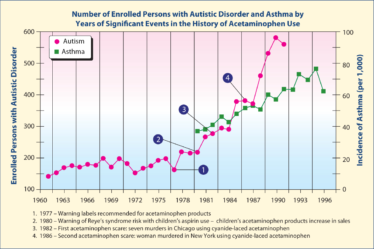
The beginning of the rapid increase in autism in around 1980 coincides with the rapid increase in asthma, both of which coincide with the rapid increase in the use of acetaminophen following the Reye's syndrome scare over a possible association with aspirin.

It would seem likely that perhaps some children on the autistic spectrum might have both brain and lung abnormalities caused by acetaminophen. Is there any evidence for such combined abnormalities? Indeed, at the annual meeting of the American College of Chest Physicians in Honolulu, Hawaii in 2011, Barbara Stewart, a pediatric pulmonologist, found that a significant lung abnormality was present in 100% of children (n = 47) on the autistic spectrum who were examined, but in none of <300 children without autism.68 Most of the children with autism had been referred to her clinic due to persistent cough unresponsive to treatment. She noticed during bronchoscopy examinations, in which a lighted tube is inserted into the lungs, that, although the airways of the children initially appeared normal, the lower airway had doubled branches, or "doublets". Dr. Stewart said "when airways divide beyond the first generation, they typically branch like a tree, with one branch on one side and one on the other. A doublet occurs when there are twin branches that come off together instead of one, which are exactly symmetrical, in each of the lower locations that can be seen." In a study of ASD children in Sweden, airway symptoms of wheezing and physician-diagnosed asthma in the baseline investigation in infants and toddlers were associated with ASD 5 years later.[52] It would seem very useful to examine the use of acetaminophen both prenatally and postnatally in the children with the abnormal lung anatomy.


DNA Damage

As of 2012, 145 studies had shown an association between mitochondria function and autism.[3] The data of Cover et al. showed that nitration of mitochondrial proteins and depletion of mitochondrial DNA after acetaminophen overdose in mice was due to peroxynitrite formation.[32] In contrast, nDNA damage was not directly caused by peroxynitrite. Nuclear DNA damage after acetaminophen overdose is likely to be caused by DNase(s) unrelated to the caspase-activated DNase, which is typically responsible for DNA fragmentation during apoptosis. The data from Cover et al. suggest that the activation of these DNase(s) is dependent on the mitochondrial oxidant stress and peroxynitrite formation.[32]

Recent data have demonstrated that nitrated tyrosine residues, as well as acetaminophen adducts, occur in the necrotic cells following toxic doses of acetaminophen. Nitrotyrosine was postulated to be mediated by peroxynitrite, a reactive nitrogen species formed by the very rapid reaction of superoxide and nitric oxide (NO). Peroxynitrite is normally detoxified by GSH, which is depleted in acetaminophen toxicity. NO synthesis (serum nitrate plus nitrite) was dramatically increased following acetaminophen.


NAPQI

The metabolism of acetaminophen (Figure 2) by CYP2E1 forms NAPQI, a reactive metabolite that binds to cysteine residues in cellular proteins and forms acetaminophen protein adducts, referred to as acetaminophen-cysteine complexes (APAP-CYS).

Heard et al. demonstrated that low concentrations of APAP-CYS are detectable in serum following therapeutic dosing with acetaminophen in the vast majority of individuals.[49] The APAP-CYS concentrations following acetaminophen overdose varied widely. Serum APAP-CYS concentrations in patients with hepatic injury following acetaminophen overdose were generally much higher than those observed during therapeutic dosing. However, three overdose patients had APAP-CYS concentrations that were of the same order of magnitude as those observed with therapeutic dosing. Adduct concentrations varied according to the degree of exposure. Currently, an absolute adduct level exceeding 1.1 µmol/L appears consistent with acetaminophen toxicity. While therapeutic dosing generally produces concentrations below 0.5 µmol/L, values in people taking recommended dosages have been reported to be very close to the lower limit of APAP-CYS concentration associated with acetaminophen toxic overdoses (1.0 µmol/L).


NAPQI and Anomalous Hair Metal Values in Autism

Concentrations of the NAPQI metabolite increased to values as high as 1.0 µmol/L or 1000 nmol/L serum following therapeutic doses of acetaminophen. Hair proteins contain a considerable amount of cysteine, and hair proteins in hair follicles and would be expected to react with NAPQI to form NAPQI adducts with cysteine sulfhydryl groups in hair. Such cysteine groups also react strongly with heavy metals such as mercury. In comparison, the mean blood mercury concentration of children with autism was 19.53 nmol/L. Thus the concentration of NAPQI might be as much as 51 times the concentration of total mercury. Since NAPQI amounts are so much higher than mercury values, it would be expected that NAPQI might significantly reduce the capacity of hair proteins to bind mercury if the binding capacity of the hair sulfhydryl groups was exceeded by the previous binding of NAPQI. Such inhibition by NAPQI may help to explain anomalous data in the measurement of mercury in hair samples of children with autism. In two baby hair studies in which samples were obtained at first haircuts, mercury values in hair were much lower in children with autism compared with those of normal controls.[69, 70] In addition, children with the most severe symptoms of autism had the lowest amount of hair mercury. Studies in Kuwait and Saudi Arabia found opposite results in children with autism; children with autism had much higher amounts of hair mercury as well as other heavy metals, but in both of these studies the children were much older (mean ages 8.8 years in Saudi Arabia and 4.2 years in Kuwait) than those in the baby hair studies (12–24 months of age).[71, 72] All of this data could be explained by patterns of acetaminophen drug use in children. Acetaminophen is commonly used prophylactically to prevent fever in infants and toddlers who receive the bulk of their vaccines in the first 2 years of life. In contrast, few vaccines and attendant prophylactic acetaminophen are administered to older children with autism in the age range in which hair mercury values were elevated compared with controls. In these older children, hair metals such as mercury would be considerably higher due to a lack of competition from NAPQI for reactive sulfhydryl groups in hair proteins in the hair follicles.


Special Concerns About the Long-Term Defective Quality Control of Acetaminophen Products

All of the information to this point has been directed to the toxicity of pure acetaminophen. Toxicity is usually due to exceeding the recommended daily dosage, often occurring due to accidental combination use of multiple products containing acetaminophen or through changes in dosage amounts by manufacturers. Adding to this are reports that acetaminophen products have been contaminated at various times, potentially increasing health risks. These problems have been documented in Food and Drug Administration inspection reports and subsequent recalls.

From the time it was introduced in 1955, acetaminophen has become one of the most successful OTC drugs, and has earned the primary manufacturer, Johnson & Johnson, an estimated US$1.3 billion every year.[73]Within the last 10 years, the FDA has chronicled manufacturing problems including mislabeling of children's tablets, where packages of the product listed an incorrect amount of the ingredients per tablet, inadequate cleaning of equipment used for manufacturing, insufficient follow-up in investigating consumer complaints, product mix-ups, and contamination with metal fragments and bacteria.[74, 75] The latter resulted in a recall of 135 million bottles of children's medications containing acetaminophen.

Additional recalls occurred in 2009 and 2010 due to trace amounts of the chemical 2,4,6-tribromoanisole found in infant, children, and adult OTC products. This recall was prompted by 775 consumer reports of nausea, vomiting, stomach pains and diarrhea received by FDA due to the contamination.[76] It was found that the chemical 2,4,6-tribromoanisole (a metabolite of a chemical fungicide tribromophenol) was being used in a manufacturing facility to treat the wood used in pallets used for transporting packaged materials.[77]Tribromoanisole is produced when naturally-occurring airborne fungi and/or bacteria (usually Aspergillus sp.,Penicillium sp., Actinomycetes, Botrytis cinerea, Rhizobium sp., or Streptomyces) are presented with brominated phenolic compounds, which they then convert into bromoanisole derivatives.

Other FDA-publicized inspection reports of acetaminophen-manufacturing facilities have listed inadequate quality controls, lack of safeguards to isolate "rejected" raw materials and other drugs, and several human errors resulting in product mix-ups.[78] Several recalls have occurred due to failed FDA manufacturing inspections and contaminated products affecting >300 million bottles of adult and children's medicines.[79, 82] Manufacturing controls are an integral aspect of pharmaceutical production to ensure public safety, and disregard or deficiency can lead to serious outcomes. In response to the high number of inadequacies found through inspections and numerous recalls, US health authorities have taken over supervision of three acetaminophen manufacturing plants to mitigate risks.[83]


A Call to Action

A large-scale, long-term study of both prenatal and postnatal effects of acetaminophen exposure on the incidence of autism, attention deficit with hyperactivity, and asthma should be conducted. However, such a study would probably take at least 5 years. If acetaminophen is indeed the cause of all of these illnesses, should acetaminophen use continue for such a long period of time while millions of additional children are further affected? Respected physicians consider that the connection of acetaminophen with asthma has been proven beyond a reasonable doubt. Dr McBride, Professor of Pediatrics at Department of Pediatrics, Northeast Ohio Medical University, Rootstown, Ohio summarizes the evidence for the acetaminophen asthma findings[45]: "There remains a possibility that confounding variables might explain some or all of the association between acetaminophen and asthma. For this reason we need further studies. At present, however, I need further studies not to prove that acetaminophen is dangerous but, rather, to prove that it is safe. Until such evidence is forthcoming, I will recommend avoidance of acetaminophen by all children with asthma or those at risk for asthma and will work to make patients, parents, and primary care providers aware of the possibility that acetaminophen is detrimental to children with asthma."

Since all children may be at risk from asthma, Dr. McBride is in effect saying that acetaminophen is contraindicated for the treatment of any children. Although the case for acetaminophen being a cause of autism and attention deficit with hyperactivity may not be as strong as the case for asthma, the severe asthma risk combined with the risks of autism and attention deficit with hyperactivity are so severe that we as a society should maintain a degree of caution with acetaminophen given the proven overall toxicity due to accidental overdose of the drug, and the availability of ibuprofen or abstaining from treatment as alternatives. A large-scale trial of acetaminophen restriction in pregnancy and the first 3 years of life is warranted to test the hypothesis that acetaminophen is a causative agent in autism, asthma, and attention deficit with hyperactivity. Due to the increased risks associated with accidental overuse of acetaminophen, increased public awareness of such risks is paramount.

Given associations of acetaminophen with increased rates of cancer, increased testicular damage, increased rates of asthma, and allergy, plausible causation of autism, and in vitro evidence of brain damage associated with metabolites of acetaminophen, new assessments of the relative risk of aspirin causing Reye's syndrome versus the risks of acetaminophen in children should be undertaken. If a clear link between acetaminophen use pre- or post-natally and autism is established, medical practice guidelines may need to be adjusted and alternative analgesics or antipyretics, such as ibuprofen, recommended. However, such a relationship may be difficult to establish, as studies may have to account for variations in dosage amounts, formats, combination product use, and for the possibility of product recalls given the recent reported manufacturing issues surrounding acetaminophen.


Note Added in Proof

After this article was submitted for publication on January 2, 2013 the following article was published prior to publication which strongly supports the hypothesis in the current article.

In 2013, Bauer and Kriebel reported that prenatal use of acetaminophen was strongly correlated with autism/Autism Spectrum Disorder prevalence (r = 0.80) using all available country-level data (n = 8) for the period 1984 to 2005. In addition, the authors found that after acetaminophen became commonly used to treat circumcision pain after 1995, there was a strong correlation between countrylevel (n = 9) autism/ASD prevalence in males and a country's circumcision rate (r = 0.98). A very similar pattern was seen among US states and when comparing the three main racial/ethnic groups in the US.[84]


Clinical References:

  1. Kim YS, Leventhal BL, Koh YJ, Fombonne E, Laska E, Lim EC, et al. Prevalence of autism spectrum disorders in a total population sample. Am J Psychiatry. 2011;168(9):904–12.

  2. Hertz-Picciotto I, Delwiche L. The rise in autism and the role of age at diagnosis. Epidemiology. 2009;20(1):84–90.

  3. Rossignol DA, Frye RE. A review of research trends in physiological abnormalities in autism spectrum disorders: Immune dysregulation, inflammation, oxidative stress, mitochondrial dysfunction and environmental toxicant exposures. Mol Psychiatry. 2012;17(4):389–401.

  4. Shaw W. The unique vulnerability of the human brain to toxic chemical exposure and the importance of toxic chemical evaluation and treatment in orthomolecular psychiatry. J Orthomol Med. 2010;25(3):125–34.

  5. Miller MT. Thalidomide embryopathy: A model for the study of congenital incomitant horizontal strabismus. Trans Am Ophthalmol Soc. 1991;89:623–74.

  6. Disability-analysis of State Reports, reviewed by the Committee on the Rights of the Child (CRC) in its Pre- Sessional Working Group, 55th Session. International Disability Alliance 2010;8.

  7. http://www.autism-society.org/about-autism/. Autism Society Website 2012. Accessed August 31, 2013.

  8. Reed G, Galindo MA. Cuba's National Immunization Program. MEDICC Review. 2008;9(1):5–7.

  9. Bäckström M, Hägg S, Mjörndal T, Dahlqvist R. Utilization pattern of metamizole in northern Sweden and risk estimates of agranulocytosis. Pharmacoepidemiol Drug Saf. 2002;11(3):239–45.

  10. Torres AR. Is fever suppression involved in the etiology of autism and neurodevelopmental disorders? BMC Pediatr. 2003;2:3–9.

  11. Schultz ST, Klonoff-Cohen HS, Wingard DL, Akshoomoff NA, Macera CA, Ji M. Acetaminophen (paracetamol) use, measles-mumps rubella vaccination, and autistic disorder: The results of a parent survey. Autism. 2008;12(3):293–307.

  12. Schultz ST. Can autism be triggered by acetaminophen activation of the endocannabinoid system? Acta Neurobiol Exp. 2010;70(2):227–31.

  13. Schultz S, Desilva M, Gu TT, Qiang M, Whang K. Effects of the analgesic acetaminophen (paracetamol) and its para-aminophenol metabolite on viability of mouse-cultured cortical neurons. Basic Clin Pharmacol Toxicol. 2011;110(2):141–4.

  14. Becker KG, Schultz ST. Similarities in features of autism and asthma and a possible link to acetaminophen use. Med Hypotheses. 2010;74(1):7–11.

  15. Posadas I, Santos P, Blanco A, Muñoz-Fernández M, Ceña V. Acetaminophen induces apoptosis in rat cortical neurons. PLoS ONE. 2010;15(12):e15360.

  16. U.S. Food and Drug Administration. For consumers – Don't double up on acetaminophen. 2013. Available at:http://www.fda.gov/ForConsumers/ConsumerUpdates/ucm336581.htm . Accessed March 19, 2013.

  17. Connor DF. Problems of overdiagnosis and overprescribing in ADHD. Psychiatric Times. 2011;28(8):1–8.

  18. Center For Disease Control. Mental health in the United States: Prevalence of diagnosis and medication treatment for attention-deficit/hyperactivity disorder – United States, 2003. MMWR. 2005;54(34):842–7.

  19. Safer DJ. Are stimulants overprescribed for youths with ADHD? Ann Clin Psychiatry. 2000;12(1):55–62.

  20. Safer D, Krager JM. Hyperactivity and inattentiveness. School assessment of stimulant treatment. Clin Pediatr. 1989;28(5):216–21.

  21. Safer DJ, Zito JM, Fine EM. Increased methylphenidate usage for attention deficit disorder in the 1990s. Pediatrics. 1996;98(6):1084–8.

  22. Safer DJ, Krager JM. The increased rate of stimulant treatment for hyperactive/inattentive students in secondary schools.Pediatrics. 1994;94(4):462–4.

  23. Safer DJ, Malever M. Stimulant treatment in Maryland Public Schools. Pediatrics. 2009;106(3):533–9.

  24. Safer DJ, Krager JM. A survey of medication treatment for hyperactive/inattentive students. J Am Med Assoc. 1988;260(15):2256–8.

  25. Mandell DS, Thompson WW, Weintraub ES, Destefano F, Blank MB. Trends in diagnosis rates for autism and ADHD at hospital discharge in the context of other psychiatric diagnoses. Psychiatr Serv. 2005;56(1): 56–62.

  26. Orlowski JP, Hanhan UA, Fiallos MR. Is aspirin a cause of Reye's syndrome? A case against. Drug Saf. 2002;25(4):225–31.

  27. Van Bever HP, Quek SC, Lim T. Aspirin, Reye syndrome, Kawasaki disease, and allergies; a reconsideration of the links. Br Med J. 2004;89(12):1178.

  28. Weiner DL. Reye Syndrome. 2011. Available at: http://EMedicine.Medscape.com. Accessed August 31, 2013.

  29. Shaw W. Possible role of lysolecithins and nonesterified fatty acids in the pathogenesis of Reye's syndrome, sudden infant death syndrome, acute pancreatitis, and diabetic ketoacidosis. Clin Chem. 1985;31(7):1109–15.

  30. Kristensen DM, Hass U, Lesné L, Lottrup G, Jacobsen PR, Desdoits-Lethimonier C, et al. Intrauterine exposure to mild analgesics is a risk factor for development of male reproductive disorders in human and rat. Human Reprod.2010;26(1):235–44.

  31. Adjei AA, Gaedigk A, Simon SD, Weinshilboum RM, Leeder JS. Interindividual variability in acetaminophen sulfation by human fetal liver: Implications for pharmacogenetic investigations of drug-induced birth defects. Birth Defects Res A Clin Mol Teratol. 2008;82(3):155–65.

  32. Cover C, Mansouri A, Knight TR, Bajt ML, Lemasters JJ, Pessayre D, et al. Peroxynitrite-induced mitochondrial and endonuclease-mediated nuclear DNA damage in acetaminophen hepatotoxicity. J Pharmacol Exp Ther. 2005;315(2):879–87.

  33. Ruepp SU, Tonge RP, Shaw J, Wallis N, Pognan F. Genomics and proteomics analysis of acetaminophen toxicity in mouse liver. Toxicol Sci. 2012;65(1):135–50.

  34. Maher JJ. DAMPs ramp up drug toxicity. J Clin Invest. 2009;119(2):246–9.

  35. Lee SS, Buters JT, Pineau T, Fernandez-Salguero P, Gonzalez FJ. Role of CYP2E1 in the hepatotoxicity of acetaminophen.J Biol Chem. 1996;271(20):12063–7.

  36. Yamaura K, Ogawa K, Yonekawa T, Nakamura T, Yano S, Ueno K. Inhibition of the antibody production by acetaminophen independent of liver injury in mice. Biol Pharm Bull. 2002;25(2):201–5.

  37. Prymula R, Siegrist CA, Chlibek R, Zemlickova H, Vackova M, Smetana J, et al. Effect of prophylactic paracetamol administration at time of vaccination on febrile reactions and antibody responses in children: Two open-label, randomised controlled trials. Lancet. 2009;374:1339–50.

  38. Homme JH, Fischer PR. Prophylactic paracetamol at the time of infant vaccination reduces the risk of fever but also reduces antibody response. Evid Based Med. 2010;15(2):50–1.

  39. Fenves AZ, Kirkpatrick III HM, Patel VV, Sweetman L, Emmett M. Increased anion gap metabolic acidosis as a result of 5-oxoproline (pyroglutamic acid): A role for acetaminophen. Clin J Am Soc Nephrol. 2006;1(3):441–7.

  40. Pitt JJ, Brown GK, Clift V, Christodoulou J. Atypical pyroglutamic aciduria: Possible role of paracetamol. J Inherit Metab Dis. 1990;13(5):755–6.

  41. Miller AL. Liver damage from chronic acetaminophen dosing is dangerous, but not the only risk. Alt Med Rev. 2009;14(4):322–3.

  42. Franciscus A, Highleyman L. Acetaminophen and your liver. HCSP Fact Sheet. 2009;2.2:1–3.

  43. Walter RB, Milano F, Brasky TM, White E. Long-term use of acetaminophen, aspirin, and other nonsteroidal anti-inflammatory drugs and risk of hematologic malignancies: results from the prospective vitamins and lifestyle (VITAL) study.J Clin Oncol. 2011;94:2424–2431.

  44. Newson RB, Shaheen SO, Chinn S, Burney PG. Paracetamol sales and atopic disease in children and adults: An ecological analysis. Eur Respir J. 2000;16(5):817–23.

  45. McBride JT. Association of acetaminophen and asthma prevalence and severity. Pediatrics. 2011;128(6):1181–5.

  46. Eneli I, Sadri K, Camargo C Jr, Barr RG. Acetaminophen and the risk of asthma: The epidemiologic and pathophysiologic evidence. Chest. 2005;127(2):604–12.

  47. Somers GF. Pharmacological properties of Thalidomide (a-phthalimido glutarimide), a new sedative hypnotic drug. Br J Pharmacol Chemother. 1960;15:111–116.

  48. Good P. Did acetaminophen provoke the autism epidemic? Alt Med Rev. 2009;14(4):364–72.

  49. Heard KJ, Green JL, James LP, Judge BS, Zolot L, Rhyee S, et al. Acetaminophen-cysteine adducts during therapeutic dosing and following overdose. BMC Gastroenterol. 2011;11:20.

  50. Krakowiak P, Walker CK, Bremer AA, Baker AS, Ozonoff S, Hansen RL, et al. Maternal metabolic conditions and risk for autism and other neurodevelopmental disorders. Pediatrics. 2012;129(5):e1121–8.

  51. Windham GC, Zhang L, Gunier R, Croen LA, Grether JK. Autism spectrum disorders in relation to distribution of hazardous air pollutants in the San Francisco Bay area. Environ Health Perspect. 2006;114(9):1438–44.

  52. Larsson M, Weiss B, Janson S, Sundell J, Bornehag CG. Associations between indoor environmental factors and parental-reported autistic spectrum disorders in children 6–8 years of age. Neurotoxicology. 2009;30(5):822–31.

  53. Kim SJ, Lee MY, Kwon do Y, Kim SY, Kim YC. Alteration in metabolism and toxicity of acetaminophen upon repeated administration in rats. J Pharmacological Sci. 2009;111(2):175–81.

  54. Alcorn J, McNamara PJ. Pharmacokinetics in the newborn. Adv Drug Deliv Rev. 2003;55(5):667–86.

  55. Högestätt ED, Jönsson BA, Ermund A, Andersson DA, Björk H, Alexander JP, et al. Conversion of acetaminophen to the bioactive N-acylphenolamine AM404 via fatty acid amide hydrolase-dependent arachidonic acid conjugation in the nervous system. J Biol Chem. 2005;280(36):31405–12.

  56. O'Reilly BA, Waring RH. Enzyme and sulphur oxidation deficiencies in autistic children with knownfood/chemical intolerances. J Orthomol Med. 1993;8(4):198–200.

  57. Alberti A, Pirrone P, Elia M, Waring RH, Romano C. Sulfation deficit in "low functioning" autistic children: A pilot study. Biol Psychiatry. 1999;46(3):420–4.

  58. Schlune A, Mayatepek E. Glutathione synthetase deficiency – An inborn error of the gamma-glutamyl cycle. J Pediatr Sci.2011;3(70):2505–9.

  59. Adams JB, Baral M, Geis E, Mitchell J, Ingram J, Hensley A, et al. The severity of autism is associated with toxic metal body burden and red blood cell glutathione levels. J Toxicol. 2009;2009:532640.

  60. Ritvo ER, Freeman BJ, Scheibel AB, Duong T, Robinson H, Guthrie D, et al. Lower Purkinje cell counts in the cerebella of four autistic subjects: Initial findings of the UCLA-NSAC autopsy research reports. Am J Psychiatry. 1986;143(7):862–6.

  61. Purves D, Augustine GJ, Fitzpatrick D, Hall WC, LaMantia AS, McNamara JO, et al. Neuroscience, 4th edn. Sunderland, MA: Sinaur Associates; 2008:432–4.

  62. Vargas DL, Nascimbene C, Krishnan C, Zimmerman AW, Pardo CA. Neuroglial activation and neuroinflammation in the brain of patients with autism. Ann Neurol. 2005;57(1):67–81.

  63. Kern JK, Jones AM. Evidence of toxicity, oxidative stress, and neuronal insult in autism. J Toxicol. Environ Health B Crit Rev. 2006;9(6):485–99.

  64. Fu X, Chen TS, Ray MB, Nagasawa HT, Williams WM. p-Aminophenol-induced hepatotoxicity in hamsters: Role of glutathione. J Biochem Mol Toxicol. 2004;18(3):154–61.

  65. Gupta S, Aggarwal S, Rashanravan B, Lee T. Th1- and Th2-like cytokines in CD4+ and CD8+ T cells in autism. J Neuroimmunol. 1998;85(1):106–9.

  66. Singh VK. Plasma increase of interleukin-12 and interferon-gamma. Pathological significance in autism. J Neuroimmunol. 1996;66(1-2):143–5.

  67. Jyonouchi H, Sun S, Le H. Proinflammatory and regulatory cytokine production associated with innate and adaptive immune responses in children with autism spectrum disorders and developmental regression. J Neuroimmunol. 2001;120(1–2):170–9.

  68. Harrison L. Autism linked to unusual shapes in lungs study shows children born with a unique airway shape may have autism. WebMD Health News. 2011. Available at: http://www.webmd.com/brain/autism/news/20111025/autism-linked-to-unusual-airwayshapes. Accessed August 31, 2013.

  69. Holmes AS, Blaxill MF, Haley BE. Reduced levels of mercury in first baby haircuts of autistic children. Int J Toxicol. 2003;22(4):277–85.

  70. Adams JB, Romdalvik J, Levine KE, Hu LW. Mercury in first-cut baby hair of children with autism versus typically-developing children. Toxicol Environ Chem. 2008;90(4):739–53.

  71. Fido A, Al-Saad S. Toxic trace elements in the hair of children with autism. Autism. 2005;9(3):290–8.

  72. Al-Ayadhi LY. Heavy metals and trace elements in hair samples of autistic children in central Saudi Arabia. Neurosciences. 2005;10(3):213–8.

  73. Easton T, Herrera S. Despite bad publicity and costly legal settlements, Johnson & Johnson refuses to put ample warnings on its Tylenol labels. Torah View 2012. Available at:https://groups.google.com/forum/#!topic/alt.med.fibromyalgia/grdcWoHo5Hw. Accessed April 26, 2012.

  74. Kavilanz P. Tylenol plant: From bad to worse. CNN Money 2010. Available at:http://money.cnn.com/2010/06/22/news/companies/tylenol_plant_inspection_history/index.htm. Accessed April 26, 2012.

  75. Kavilanz P. Tylenol plant still plagued by FDA violations. CNN Money 2012. Available at:http://money.cnn.com/2010/12/01/news/companies/tylenol_plant_new_problems/index.htm. Accessed April 26, 2012.

  76. Kavilanz P. Tylenol recall: Serious side effects investigated. CNN Money 2010. Available at:http://money.cnn.com/2010/05/25/news/companies/tylenol_recall_adverse_consumer_complaints/index.htm. Accessed April 26, 2012.

  77. Kavilanz P. New recalled Tylenol linked to shut plant. CNN Money 2010. Available at:http://money.cnn.com/2010/10/19/news/companies/new_tylenol_recall/index.htm. Accessed April 26, 2012.

  78. Baker SL. Drugs like Tylenol can be contaminated with mold and chemicals. NaturalNews Com 2010. Available at:http://www.naturalnews.com/027965_Tylenol_recall.html. Accessed April 26, 2012.

  79. Heavey S. FDA finds grime at J&J plant, urges use of generics. Reuters 2010. Available at:http://www.reuters.com/article/2010/05/05/us-johnsonandjohnson-recall-idUSTRE64367Z20100505. Accessed April 26, 2012.

  80. Silverman E. Which Drugmaker Fails Most FDA Inspections? Pharmalot com 2011. Available at: https://seekingalpha.com/article/256209-which-drugmaker-fails-most-fda-inspections. Accessed April 26, 2012.

  81. Krauskopf L. More Tylenol Recalled. The Huffington Post 2011. Available at:http://www.huffingtonpost.com/2011/03/30/tylenol-recalled-_n_842450.html. Accessed April 27, 2012.

  82. Kavilanz P. U.S. takes over three Tylenol plants. CNN Money 2011. Available at:http://money.cnn.com/2011/03/10/news/companies/johnson_mcneil_fda_action/index.htm. Accessed April 27, 2012.

  83. Bauer AZ, Kriebel D. Prenatal and perinatal analgesic exposure and autism: an ecological link. Environ Health. 2013;12:41. doi: 10.1186/1476-069X-12-41.

Usefulness of HPHPA marker in a wide range of neurological, gastrointestinal, and psychiatric disorders

William Shaw, Ph.D.

The dysbiosis marker 3-(3-hydroxyphenyl)-3-hydroxypropionic acid (HPHPA), the predominant dihydroxyphenylpropionic acid isomer in urine, is also measured in the Organic Acids Test offered by The Great Plains Laboratory. This marker was proven by Dr. William Shaw to be due to a combination of human metabolism and the metabolism by a group of Clostridia species, including but not limited to C. difficile. 

HPHPA has been one of the most useful clinical markers in recent medical history. Treatment with metronidazole, vancomycin, or high doses of probiotics of individuals with high urinary values has led to significant clinical improvements or remissions of psychosis.

The biochemical role of Clostridia in altering brain neurotransmitters is due to the fact that Clostridia metabolites inactivate dopamine beta-hydroxylase, leading to an excess production of brain dopamine and reduced levels of the neurotransmitter norepinephrine. Excess dopamine is associated with abnormal or psychotic behavior. This imbalance can be demonstrated in the Organic Acids Urine Test by observing the ratio of the major dopamine metabolite, homovanillic acid (HVA), to that of the major norepinephrine metabolite, vanillylmandelic acid (VMA) when the Clostridia marker HPHPA is elevated. After treatment with metronidazole or vancomycin, HPHPA values return to normal along with normal ratios of HVA/VMA and normal behavior. 

The highest value of HPHPA was measured in the urine of a young woman with first onset of schizophrenia. Treatment of Clostridia bacteria resulted in loss of auditory hallucinations. In autism, children with gastrointestinal Clostridia commonly exhibit aggressive behavior, agitation, obsessive compulsive behavior, and irritability. They may have very foul stools with diarrhea with mucus in the stools although some individuals may be constipated. Stool testing for Clostridia is usually of limited usefulness since most Clostridia species are considered probiotics or beneficial. There are about 100 species of Clostridia that are commonly found in the gastrointestinal tract. Only seven of these species are producers of HPHPA including C. sporogenes, C.botulinum, C. caloritolerans, C. angenoti, C. ghoni, C.bifermentans, C. difficile, and C. sordellii while C. tetani,C. sticklandii, C. lituseburense, C. subterminale, C.putifaciens, C. propionicum, C. malenomenatum, C.limosum, C. lentoputrescens, C. tetanomorphum, C.coclearium, C. histolyticum, C. aminovalericum, and C.sporospheroides do not produce compounds that are converted to HPHPA.

The same article by Dr. Shaw indicates that 3,4-dihydroxyphenylpropionic acid (DHPPA) is a marker for beneficial bacteria in the gastrointestinal tract such as Lactobacilli, Bifidobacteria, and E. coli. The exception is one species of Clostridia orbiscindens that can convert the flavanoids luteolin and eriodictyol, that occur only in a relatively small food group that includes parsley, thyme, celery, and sweet red pepper to 3,4-dihydroxyphenylpropionic acid. The quantity of C. orbiscindens in the gastrointestinal tract is negligible (approximately 0.1% of the total bacteria) compared to the predominant flora of Lactobacilli, Bifidobacteria, and E. coli (7). DHPPA is an antioxidant that lowers cholesterol, reduces proinflammatory cytokines, and protects against pathogenic bacteria. 2,3-Dihydroxyphenypropionic acid, a different isomer has been claimed to be a metabolite of Pseudomonas species but the literature indicates that this compound is formed by the in vitro action of these species on quinoline, a component of coal tar, a substance missing from the diet of virtually all humans. 

References:

1. Shaw W. Increased urinary excretion of a 3-(3-hydroxyphenyl)-3-hydroxypropionic acid (HPHPA), an abnormal phenylalanine metabolite of Clostridia spp. in the gastrointestinal tract, in urine samples from patients with autism and schizophrenia. Nutr Neurosci. 2010 Jun;13(3):135-43.